Physical and psychophysical evaluation of digital systems for radiology
The purpose of this research is to evaluate the performance of digital detectors used in radiology. To assess the image quality of a digital detector, spatial resolution and noise properties can be evaluated, using metrics such as the modulation transfer function (MTF), normalized noise power spectra (NNPS), noise equivalent quanta (NEQ),and detective quantum efficiency (DQE). Such parameters have the potential for providing objective assessments of imaging performance for an image as viewed by an ideal observer. Measurements of this type allow objective comparisons to be made between different systems. A software which aims to assist users in achieving such physical characterization can be downloaded here.
Image quality of a digital detector may be characterized using these objective measures, however medical diagnosis also involves the perception by the observer. Images are a means for visually representing the clinical information captured by the FFDM units. Consequently, it is expected that image quality should be based on the judgment of a human observer. Contrast detail (CD) phantoms are often used for evaluating the image performance of a system, by involving human observers. These phantoms consist in a number of objects with different size and thickness. They are used for individuating the boundary between visible and invisible objects acquired by the detectors. CD analysis is a step towards the examination of details resembling clinical lesions, even if results obtained with CD studies could not be directly extended to clinical detection tasks. Two of the most common phantoms used in mammography and radiology are CDMAM and CDRAD, respectively.
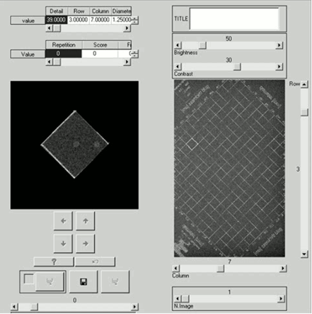
The reading of CD phantoms by human observers is a very tedious and time consuming task. We developed dedicated software, for facilitating the CDMAM reading by human observers. The software is written in IDL language, and incorporates all the features of the CDMAM phantom (sizes, thicknesses, and positions of all the details). Observers can select which square within the phantom they want to visualize and choose the location (vertex of the square) where the eccentric gold disk is supposed to be. Crops are randomly rotated by an angle of 90, 180, or 270 degrees to prevent any "memory effects" in the readers about the location of the disks within each square. When several phantom images are acquired in the same acquisition conditions, the software visualize in a random order the corresponding squares coming from the different realizations. The following picture shows the main panel of this software. The software can be downloaded here.

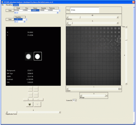
Besides, high inter and intra observer variability can affect the effectiveness of the subjective observations obtained with CD analysis. To surpass this shortcoming, automatic methods have been developed and evaluated. The use of an automatic technique can thus help to reduce the observer subjective variability, and the time required for analyzing several images dramatically decreases. We developed a software that automatically reads the CDRAD images. After a manual registration of the phantom, the software scans all the cells of the phantom and calculates the SNR for the central detail within the cells. The following picture shows the main panel of this software. The software can also be used for assisting a reading by human observers. The software can be downloaded here.


